Treatment in manifestation of hemangioma in oral cavity: A review article
Nanda Rachmad Putra Gofur1*, Aisyah Rachmadani Putri Gofur2, Soesilaningtyas3, Rizki Nur Rachman Putra Gofur4, Mega Kahdina4, Hernalia Martadila Putri4
1 Department of Health, Faculty of Vocational Studies, Universitas Airlangga, Surabaya, Indonesia
2 Faculty of Dental Medicine, Universitas Airlangga, Surabaya, Indonesia
3 Department of Dental Nursing, Poltekkes Kemenkes, Surabaya, Indonesia
4 Faculty of Medicine, Universitas Airlangga, Surabaya, Indonesia
Abstract
Background: Hemangioma is the most common benign vasoformative tumors of infancy and childhood. At least 10–20% cases needs active intervention because of their tendency to bleed and become ulcerated. Hemangiomas of the oral cavity are not common but amongst them, the head and neck is the common site. Incidence of Hemangioma ranges from 1–12% depending on age and population. Objective: To know the management of hemangioma in oral cavity. Problem Statement: There are an increasing case of hemangioma, and it is usually manifested in oral cavity. This can become bigger problem in human body. So we have to know the management of hemangioma in oral cavity to prevent it.
Discussion: The diagnoses of hemangioma are s from the history and clinical examination. Lesion shich is superficial and localized such as one in the tongue, imaging study is usually not indicated. If the lesion is accessible surgically, surgical excision is the gold standard treatment.
Conclusion: There are several treatment of hemangioma in oral cavity. But, sclerosing agent with 3% sodium tetradecyl sulfate is very effective for treatment of oral hemangioma with the certain dosage and the site of injection according to the sive of lesion so the complications can be prevent, after that laser or surgery is needed.
Keywords: oral hemangioma, surgery, oral hemangioma treatment
Introduction
Haemangioma are the most common benign vasoformative tumours of infancy and childhood. They usually are manifested within the first month of life, exhibit a rapid proliferative phase, and slowly involute to near complete resolution. There are many ways to classify haemangiomas. Haemangiomas are broadly classified into capillary, cavernous, and miscellaneous forms like verrucous, venous, arteriovenous haemangiomas, and so forth. Capillary haemangiomas further include juvenile, pyogenic granuloma, and epitheliod haemangioma. The term haemangioma has been commonly misused to describe a large number of vasoformative tumours [1, 2].
Nowadays there are so many disease which can occur in vascular, and it can become problem in human body. One of the vascular disease which can become the problem is hemangioma. Hemangioma is the most common benign vasoformative tumors of infancy and childhood. Hemangioma usually is manifested in the first month of life, it exhibits a rapid proliferative phase, and slowly involute to near complete resolution. At least 10–20% cases needs active intervention because of their tendency to bleed and become ulcerated. The benign, localized tumor of the blood vessels is called as hemangioma. Most of the benign vascular lesions occurring in the head and neck region usually have a malformation, hamartomatous basis [3, 4].
Hemangioma can manifested in oral cavity. Hemangiomas of the oral cavity are not common but amongst them, the head and neck is the common site. Incidence of Hemangioma ranges from 1–12% depending on age and population. Injection of sclerosing agents into these lesions can serve as therapy as well as a preoperative treatment. The common place of haemangioma involve the head and neck. However, they are rare in the oral cavity but may occur on tongue, lips, buccal mucosa, gingiva, palatal mucosa, salivary glands, alveolar ridge, and jaw bones. Management of haemangioma depends on size, exact location, stages of growth or regeneration, functional compromise, and behaviour [3, 4]. Aim of this study is to know the management of hemangioma in oral cavity [5, 6].
Discussion
Hemangiomas of the oral cavity are not common pathologic entities, but, among hemangiomas, the head and the neck are common sites. Most true hemangiomas involute with time, but a certain small percentage do not, which may present with complications that require treatment (see Complications). An estimated 10-20% of true hemangiomas incompletely involute and require postadolescent ablative treatment [7].
Pathogenesis and origin of haemangioma remain incompletely understood. However, various theories have been proposed to elucidate the mechanism and pathogenesis of haemangioma. Aberrant and focal proliferation of endothelial cells results in haemangioma, although the cause behind this remains unclear. The placental theory of haemangioma origin has been described who studied various histology and molecular markers such as GLUTI, Lewis Y Antigen, Merosin, CCR6, CD15, IDO, FC, and gamma Receptor II [9, 8].
Clinically, haemangioma appears as soft mass, smooth or lobulated, and sessile or pedunculated and may vary in size from a few mms to several cms. They are usually deep red and may blanch on the application of pressure and if large in size, might interfere with mastication. In the present case study, there is a rare and an unusual case of capillary haemangioma of the palatal mucosa [10, 11].

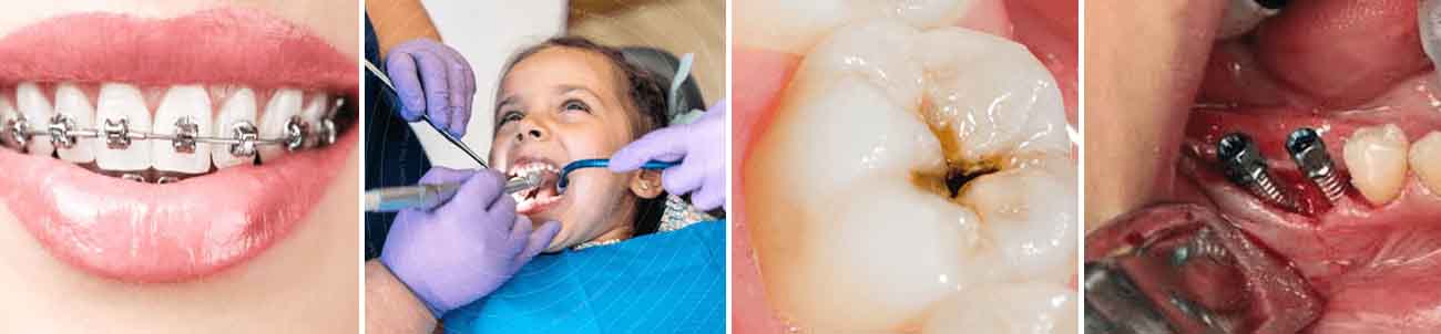
Fig 1: Clinical Manifestation Oral Hemangioma in Mucosa and Toungep [11].
Hemangiomas are usually classified into capillary, cavernous and mixed hemangioma. In oral cavity the bone and muscles are affected as well as the mucosa and skin. The diagnoses of hemangioma are s from the history and clinical examination. Lesion shich is superficial and localized such as one in the tongue, imaging study is usually not indicated. If the lesion is accessible surgically, surgical excision is the gold standard treatment. However there are several obstacles when considering surgery such as: 1) Complete excision is not possible; 2) Dissection is often complicated by excessive bleeding; 3) Recurrence; 4) Functional impairment of vital functions like swallowing; 5) Morbidity of surgical procedure; These issues have led people to seek alternative treatment of these malformations like cautery, cryotherapy, radiotherapy, sclerosing agents. Sclerosing agent causes marked tissue irritation and/or thrombosis with subsequent local inflammation and tissue necrosis. The inflammation and tissue necrosis result in fibrosis with tissue contracture. Sodium tetradecyl sulfate (sotradecol) is the sclerosing agent which has been used for years in the treatment of varicose vein, hemorrhoids and hemangioma [12, 13, 14].
Use of laser therapy for the treatment of hemangioma has gained popularity. Laser leads to selective photothermolysis rather than non-selective tissue destruction. Oral mucosa can be amenable to yellow light laser (578–585 nm) as these are selectively absorbed by hemoglobin. The tunable dye laser can ablate superficial ecstatic blood vessels without significant epidermal damage or scarring [15, 16].
Management of haemangioma depends on a variety of factors, and most true haemangioma requires no intervention. However, 10–20% requires treatment because of the size, exact location, stages of growth or regeneration, functional compromise, and behaviour. The range of treatment includes surgery, flash lamp pulsed laser, intralesional injection of fibrosing agent, interferon alpha-2b, and electrocoagulation while cryosurgery, compression and radiation were used in the past. Each treatment modality has its own risk and benefits [17, 18].
Conclusion
There are several treatment of hemangioma in oral cavity. But, sclerosing agent with 3% sodium tetradecyl sulfate is very effective for treatment of oral hemangioma with the certain dosage and the site of injection according to the sive of lesion so the complications can be prevent, after that laser or surgery is needed.
References
- Singh P, Parihar AS, Siddique SN, et al. Capillary haemangioma on the palate: a diagnostic conundrum. BMJ Case Rep, 2016, 16.:pii: bcr2015210948.
- Hydoh H, Hori M. Periphral vascular malformations, imaging, treatment approaches and therapeutic issues. Radiographics 25(special issue), 2005, 159-172.
- Radhika BN, Lankupalli AS. Lesions of Lip and Tongue. IOSR Journal of Dental and Medical Sciences (IOSR-JDMS). 2014; 13(2):1-5.
- Buckmiller LM, Richter GT, Suen JY. “Diagnosis and management of hemangiomas and vascular malformations of the head and neck,” Oral Diseases. 2010; 16(5):405-18.
- Qiam F, Khan M, Din QU. Mucosal venous hemangiomas of the oral cavity – an analysis of 43 cases and literature review. JKCD. 2011; 2(1):1-5.
- Buckmiller LM, Richter GT, Suen JY. Diagnosis and management of hemangiomas and vascular malformations of the head and neck. Oral Diseases. 2010; 16(5):405-418.
- North PE, Waner M, Brodsky MC. Are infantile hemangiomas of placental origin? Ophthalmology. 2002; 109(4):633-634.
- Kleinman ME, Greives MR, Churgin SS et al. Hypoxia-induced mediators of stem/progenitor cell trafficking are increased in children with hemangioma. Arteriosclerosis, Thrombosis, and Vascular Biology. 2007; 27(12):2664-2670.
- Frischer JS, Huang J, Serur A, Kadenhe A, Yamashiro DJ, Kandel JJ. Biomolecular Markers and Involution of Hemangiomas. Journal of Pediatric Surgery. 2004; 39(3):40 0-404.
- Sun ZJ, Zhao YF, Zhao JH. Mast cells in hemangioma: a double-edged sword. Medical Hypotheses. 2007; 68(4):805-807.
- Portaro, Simona MD, PhDa; Naro, Antonino MD, PhDa; Guarneri, Claudio MDb; Di Toro, Giuseppe MDc; Manuli, Alfredo Msca; Calabrò, Rocco Salvatore MD, PhDa,* Hemangiomas of the tongue and the oral cavity in a myotonic dystrophy type 1 patient, Medicine: November 2018; 97(48):13448 doi: 10.1097/MD.0000000000013448
- Burstein FD, Simms C, Cohen SR, Williams JK, Paschal M. Intralesional laser therapy of extensive hemangiomas in 100 consecutive pediatric patients. Annals of Plastic Surgery. 2000; 44(2):188-194.
- Onesti MG, Mazzocchi M, Mezzana P, Scuderi N. Different types of embolization before surgical excision of haemangiomas of the face. Acta Chirurgiae Plasticae. 2003; 45(2):55-60.
- Bonet-Coloma C, Mínguez-Martínez I, Palma-Carrió C, et al. Clinical characteristics, treatment and outcome of 28 oral hemangiomas in pediatric patients. Med Oral Patol Oral Cir Bucal. 2011; 16:e19-22.
- Avila ED, Molon RS, Conte Neto N et al. Lip cavernous hemangioma in a young child. Braz Dent J. 2010; 21:370-4.
- Slaba S, Braidy C, Sader RB, et al. Giant venous malformation of the tongue: the value of surgiflo. J Mal Vasc. 2010; 35:197-201.
- Atkins JH, Mandel JE, Mirza N. Laser ablation of a large tongue hemangioma with remifentanil nalgosedation in the ORL endoscopy suite. ORL J Otorhinolaryngol Relat Spec. 2011; 73:166-9.
- Kutluhan A, Bozdemir K, Ugras S. The treatment of tongue haemangioma by plasma knife surgery. Singapore Med J. 2008; 49:e312-4.
Please enter the email address corresponding to this article submission to download your certificate.